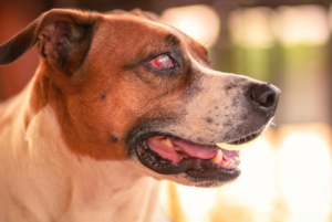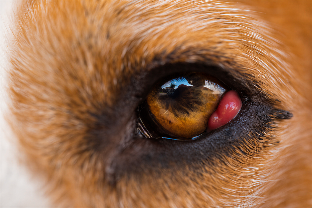
Cherry eye is a prolapse of the tear gland in a pet’s third eyelid. It looks like a smooth, red, swollen mass in the inner corner of the eye, near the nose. It is not a cancerous mass and usually is not painful at first, but it should be treated with surgery that repositions—not removes—the gland, because that gland produces much of the eye’s tears. It is most common in young dogs and certain breeds, and it will not go back into place on its own. Did you spot a small red bubble in the corner of your dog’s eye? Here’s what it is, why it happens, and how we treat it at Visionary Vet Services in Fort Myers.
What is cherry eye?
Dogs and cats have a third eyelid, a protective membrane in the inner corner of the eye, and tucked at its base is a tear-producing gland. Cherry eye is the common name for prolapse of that gland: it slips out of position and pops into view as a red, fleshy lump resembling a cherry. This gland is not optional; it produces a large share of the tear film’s watery layer, which drives the modern treatment approach.
What does cherry eye look like?
The classic sign is a smooth, rounded, pink-to-red mass bulging from the inner corner of the eye nearest to the nose. It can appear suddenly, sometimes overnight, and affect one or both eyes. Many pets show no discomfort at first, but over time, the exposed gland can dry out and become inflamed, leading to redness, discharge, rubbing, reduced tear function, and conjunctivitis.
What causes cherry eye?
Cherry eye is thought to come from a weakness in the connective tissue that anchors the gland in place. There is a strong genetic and breed component: a 2024 genome-wide study linked cherry eye to a specific genetic variant and confirmed it is largely heritable. Breeds seen most often include:
- English and French Bulldogs
- Cocker Spaniels and Beagles
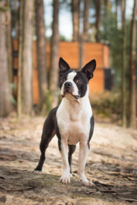
- Boston Terriers
- Shih Tzus
- Great Danes
Most cases appear in dogs under two years old. Cats can be affected, though rarely. If one eye prolapses, the other often follows, something we discuss at the exam.
Why shouldn’t the gland just be removed?
This is the most important point. The gland used to be cut out; we now know that is a mistake. Research shows the prolapsed gland is still functional tissue worth preserving. Removing it sharply raises the risk of dry eye (keratoconjunctivitis sicca) later, a chronic, uncomfortable condition requiring lifelong daily medication. The standard of care is to reposition the gland, tucking it back into place so it continues to function.
How is cherry eye treated?
Surgery is the recommended treatment and should be done soon after the prolapse appears, while the gland is healthy. The most common technique is the “pocket” method, in which the surgeon creates a small pocket in the third eyelid and sutures the gland back beneath the membrane. Success rates are high. Recurrence can occasionally occur and may require a second procedure, with some breeds (such as English Bulldogs) more prone to recurrence. Anti-inflammatory medication may calm a very early case, but it does not fix the underlying problem. Because protecting gland function requires precise technique, this surgery is well-suited to a veterinary ophthalmologist.
FAQs
- Is cherry eye painful? Not usually at first, but the exposed gland can become dry and inflamed over time.
- Will it go away on its own? No. The gland will not reliably return to its position without surgery.
- Is it an emergency? Not a same-hour emergency like glaucoma, but it should be addressed within days to a couple of weeks to protect tear production.
- Can both eyes be affected? Yes—and a dog affected in one eye is more likely to develop it in the other.
Schedule a cherry eye consultation
If your dog or cat has a red lump in the corner of the eye, early treatment gives the best chance of preserving healthy tear production for life. Contact Visionary Vet Services in Fort Myers to schedule an evaluation.
References
- Freyer J, Labadie JD, Huff JT, et al. Association of FGF4L1 retrogene insertion with prolapsed gland of the nictitans (cherry eye) in dogs. Genes (Basel). 2024;15(2):198. doi:10.3390/genes15020198
- Dugan SJ, Severin GA, Hungerford LL, Whiteley HE, Roberts SM. Clinical and histologic evaluation of the prolapsed third eyelid gland in dogs. J Am Vet Med Assoc. 1992;201(12):1861-1867. doi:10.2460/javma.1992.201.12.1861
- Mazzucchelli S, Vaillant M, Wéverberg F, et al. Retrospective study of 155 cases of prolapse of the nictitating membrane gland in dogs. Vet Rec. 2012;170(17):443. doi:10.1136/vr.100587