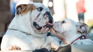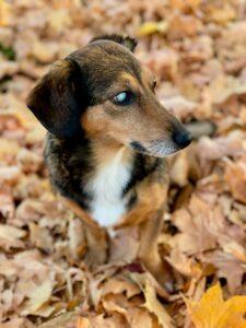
If you have a Frenchie or Bulldog in Southwest Florida, there’s a good chance you’ve either seen cherry eyes, or you will.
That little pink bubble in the corner of the eye? It’s super common in these breeds. Now let’s break it down.
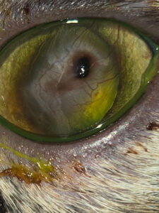
What Is Cherry Eye?
Cherry eye is a prolapse of the gland of the third eyelid. This gland normally stays tucked away but produces up to 35–50% of your dog’s tears. When it pops out of place, it creates that round, red swelling in the inner corner of the eye.¹
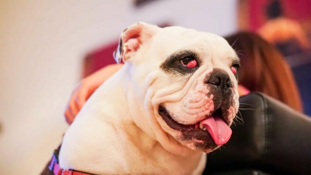
Why Are French Bulldogs and English Bulldogs So Prone?
Frenchies and Bulldogs are flat-faced breeds, so their eye anatomy makes them more likely to develop cherry eye. In the Florida heat and humidity, exposed gland tissue can also become irritated quickly, which can worsen inflammation.²
Can Cherry Eye Go Away on Its Own?
Rarely. You might see temporary improvement with anti-inflammatory drops, but the gland typically prolapses again. Cherry eye is considered a structural issue, not just inflammation.
Should the Gland Be Removed?
No—this is important. That gland plays a major role in tear production. Studies show removing it significantly increases the risk of dry eye later in life.³ And Frenchies and Bulldogs are already prone to eye surface disease. We don’t want to reduce tear production further.
What Is the Best Treatment?
Surgical repositioning. Today’s veterinary ophthalmology focuses on gently tucking the gland back into its normal position, techniques that preserve the gland, and protect long-term tear function.⁴ Most dogs recover quickly and are back to their goofy selves within a few weeks.
Is Cherry Eye an Emergency?
Not usually, but don’t ignore it. Early correction improves surgical success and reduces the risk of chronic irritation or secondary dry eye.
The Bottom Line for Florida Frenchie & Bulldog Owners
Even though cherry eye is extremely common in these breeds, it’s treatable, and outcomes are excellent when managed properly.
If you notice that pink bump in your French Bulldog or English Bulldog, schedule an appointment with us sooner rather than later. In our sunny Southwest Florida climate, protecting tear production is key to long-term eye health, and overall health, too.
References
- Gelatt KN, et al. Veterinary Ophthalmology. 5th ed. Wiley-Blackwell; 2013.
- Morgan RV, Duddy JM, McClurg K. Prolapse of the gland of the third eyelid in dogs: 89 cases. J Am Anim Hosp Assoc. 1993;29:56-60.
- Helper LC. The tear film in dogs and cats. Vet Clin North Am Small Anim Pract. 1990;20(3):617-625.
- Stanley RG, Kaswan RL. Surgical repositioning techniques for prolapsed nictitans gland. Vet Ophthalmol.